Astaxanthinアスタキサンチン
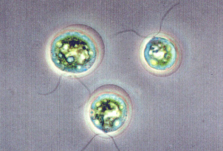
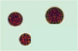
ハワイ島コナ・ケアホレポントが造る独特の自然環境が、閉鎖循環式と屋外流路式の併用による清浄なヘマトコッカス藻を育成し、
アスタキサンチンの経済生産を実現させました。


Astaxanthin Prevents Atrophy in Slow Muscle Fibers by Inhibiting Mitochondrial Reactive Oxygen Species via a Mitochondria-Mediated Apoptosis Pathway
Luchuanyang Sun1,Nobuyuki Miyaji2,Min Yang1,Edward M. Mills3,Shigeto Taniyama1,Takayuki Uchida4,Takeshi Nikawa4,Jifeng Li5,Jie Shi5,Katsuyasu Tachibana1 and Katsuya Hirasaka16
1 Graduate School of Fisheries and Environmental Sciences, Nagasaki University, Nagasaki 8528521, Japan
2 Toyo Koso Kagaku Co., Ltd., Chiba 2790041, Japan
3 Division of Pharmacology/Toxicology, University of Texas at Austin, Austin, TX 78712, USA
4 Department of Nutritional Physiology, Institute of Medical Nutrition, Tokushima University Medical School, Tokushima 7708503, Japan
5 Weihai Lida Biological Technology Co., Ltd., Weihai 264200, China
6 Organization for Marine Science and Technology, Nagasaki University, Nagasaki 8528521, Japan
Author to whom correspondence should be addressed.
Nutrients 2021, 13(2), 379; https://doi.org/10.3390/nu13020379
Received: 17 June 2020 / Revised: 19 January 2021 / Accepted: 22 January 2021 / Published: 26 January 2021
(This article belongs to the Special Issue Dietary Antioxidants for Human Health)
Download PDF Browse Figures Review Reports Citation Export
Abstract
Astaxanthin (AX) is a carotenoid that exerts potent antioxidant activity and acts in the lipid bilayer. This study aimed to investigate the effects of AX on muscle-atrophy-mediated disturbance of mitochondria, which have a lipid bilayer. Tail suspension was used to establish a muscle-atrophied mouse model. AX diet fed to tail-suspension mice prevented loss of muscle weight, inhibited the decrease of myofiber size, and restrained the increase of hydrogen peroxide (H2O2) production in the soleus muscle. Additionally, AX improved downregulation of mitochondrial respiratory chain complexes I and III in the soleus muscle after tail suspension. Meanwhile, AX promoted mitochondrial biogenesis by upregulating the expressions of adenosine 5′-monophosphate?activated protein kinase (AMPK) α-1, peroxisome proliferator?activated receptor (PPAR)-γ, and creatine kinase in mitochondrial (Ckmt) 2 in the soleus muscle of tail-suspension mice. To confirm the AX phenotype in the soleus muscle, we examined its effects on mitochondria using Sol8 myotubes derived from the soleus muscle. We found that AX was preferentially detected in the mitochondrial fraction; it significantly suppressed mitochondrial reactive oxygen species (ROS) production in Sol8 myotubes. Moreover, AX inhibited the activation of caspase 3 via inhibiting the release of cytochrome c into the cytosol in antimycin A?treated Sol8 myotubes. These results suggested that AX protected the functional stability of mitochondria, alleviated mitochondrial oxidative stress and mitochondria-mediated apoptosis, and thus, prevented muscle atrophy.